
HARUE DENTAL · FULL-MOUTH IMPLANTS
When an existing implant-supported restoration fails, I first assess the remaining implants, bone and nerve positions. This patient had difficulty eating after part of an upper restoration came out with implants. Peri-implantitis was also present around lower molar implants.
Treatment and explanation: Dr Hanna Lee

Photographed: 2024.09.10
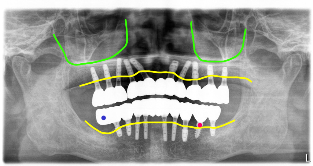
Photographed: 2025.03.21
Case overview
- Revision planned for peri-implantitis, bone loss and a failed restoration
- Same-day fixed temporary teeth; about six months and four visits
- A serviceable existing lower implant was retained
01 · Planning revision treatment
I recommended removing implants that were unlikely to remain serviceable, then placing implants using the remaining bone. The patient had developed dental anxiety after severe gagging during earlier surgery. Sedation was prepared, but on the day the patient felt comfortable and treatment proceeded without it. This experience does not predict another patient’s discomfort or sedation needs.
02 · Assessing bone and nerve positions
In the upper jaw, I removed four existing implants affected by peri-implantitis and placed ten implants in the remaining bone. In the lower jaw, I retained a serviceable existing implant and planned the new implants around the available bone and nerve position.
I avoided placement in the lower right posterior region because of severe inflammation and proximity to the nerve canal. A cantilever extension was used for that region. One existing implant on the lower left with a favourable condition was retained. I also adjusted the reduced bite height. Cantilever suitability and loading must be assessed individually.
03 · Temporary teeth and the healing period
Fixed temporary teeth were fitted on the day of surgery. Treatment was completed over about six months and four visits. Same-day temporary teeth and the later definitive restorations are separate stages.
This patient followed a liquid diet while waiting for adequate implant stability. Diet during healing should follow the treating clinician’s instructions. The earlier record notes regular follow-up, but does not establish the patient’s current condition.
The images are dated 10 September 2024 before revision and 21 March 2025 at post-treatment follow-up. The latter is a photograph date, not confirmation of the exact date of final restoration placement.