
1. Missing Front Teeth: Is Filling the Space Enough?
When a front tooth is missing, we first assess the remaining tooth positions and spaces. In this case, both lateral incisors were absent. Existing resin was removed, orthodontics redistributed the spaces, and veneers adjusted the shapes of the front teeth and canines.
This is a missing front-tooth case with both lateral incisors absent. Orthodontics repositioned the spaced teeth before veneers refined their contours.
A missing tooth describes the absence of a tooth. Here, the missing front teeth were those immediately beside the large central incisors on both sides: bilateral missing lateral incisors. Missing lateral incisors may leave spaces or allow canines to move forward. Before filling every gap with restorations, we assess widths, proportions, the bite and the relationship of gums and lips within the smile.
Initial condition
Both lateral incisors absent, front-tooth spacing and existing resin restorations
Treatment sequence
Resin removal → orthodontic space redistribution → veneers
Treatment focus
Coordinating central-incisor widths and canine contours
Photo record
2015.08.04 / 2016.01.12 / 2016.02.19
2. Before and After at a Glance
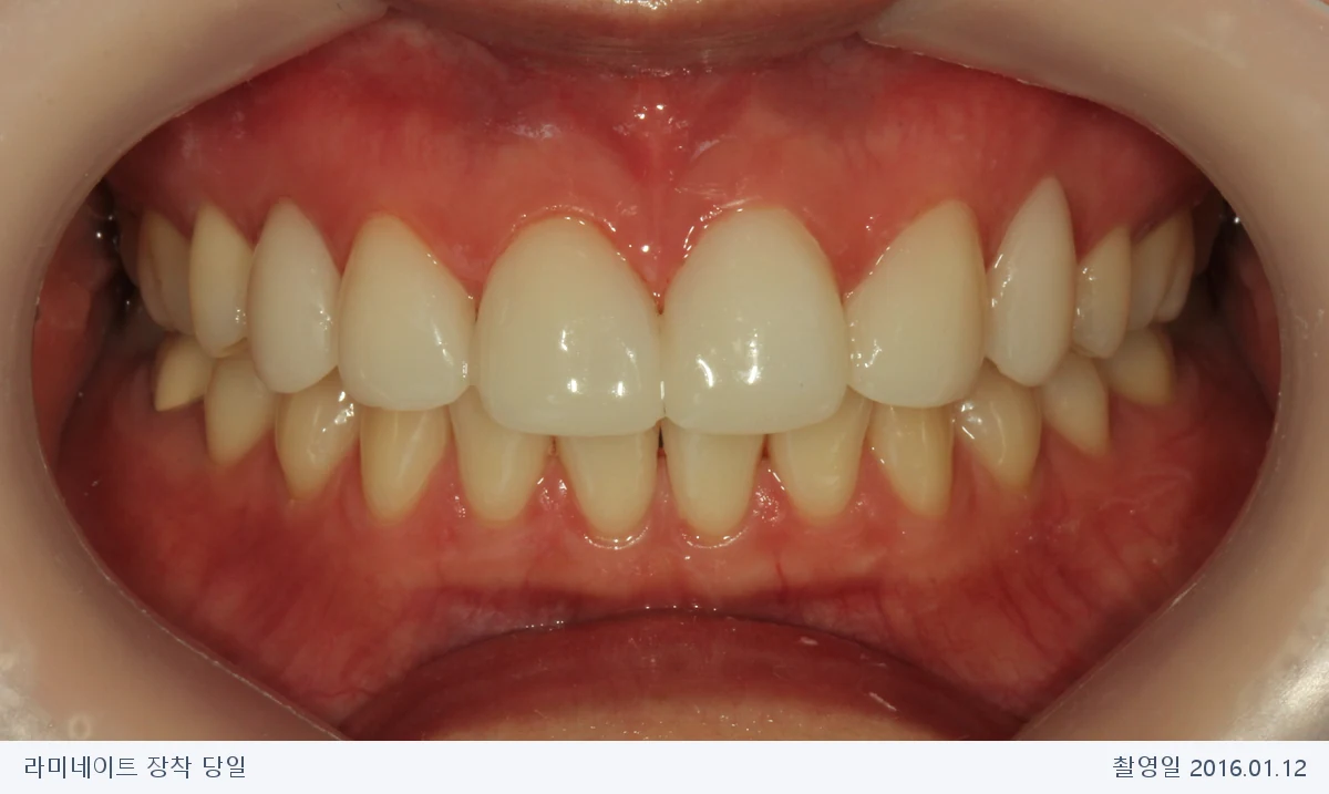
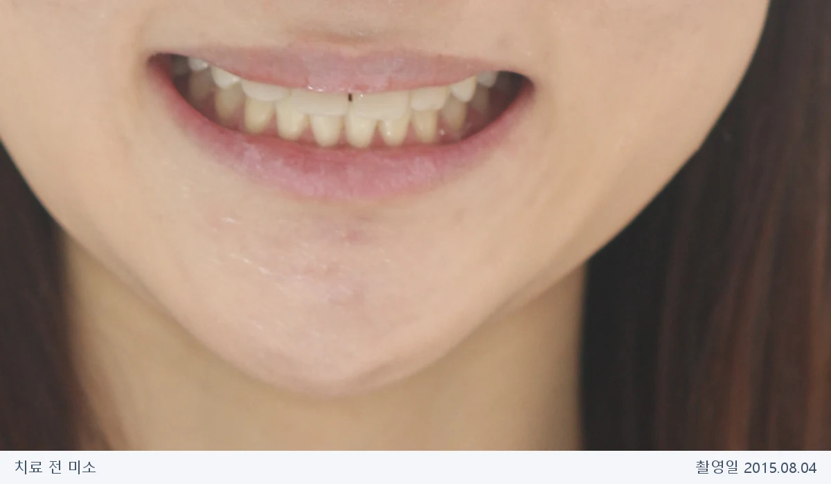
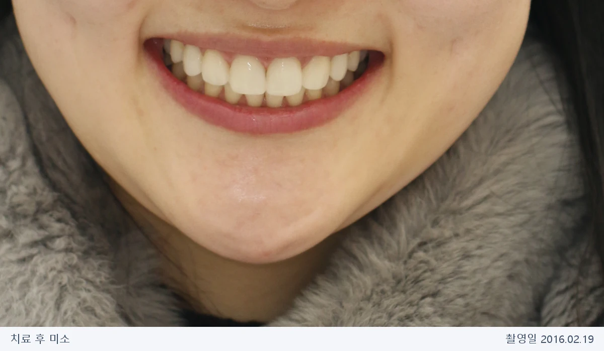
Frontal photographs of the same patient treated at Harue Dental. The left shows the old resin before treatment; the right is the veneer fitting day after orthodontics. The result reflects both treatments together.
Results vary between patients. Tooth colors and contours were not retouched; lighting and angles differ between dates.
Key risks and precautions: Orthodontics may cause pain, decay, gum changes, root resorption and relapse. Veneers may require irreversible preparation; fracture, debonding or surrounding decay can lead to retreatment.

3. Questions You May Have
Do missing front teeth always require implants?
Why not close the spaces directly with veneers?
What if a gap returns after resin bonding?
These are general questions to help explain treatment choices.
4. Why Was Orthodontics Done before Veneers?
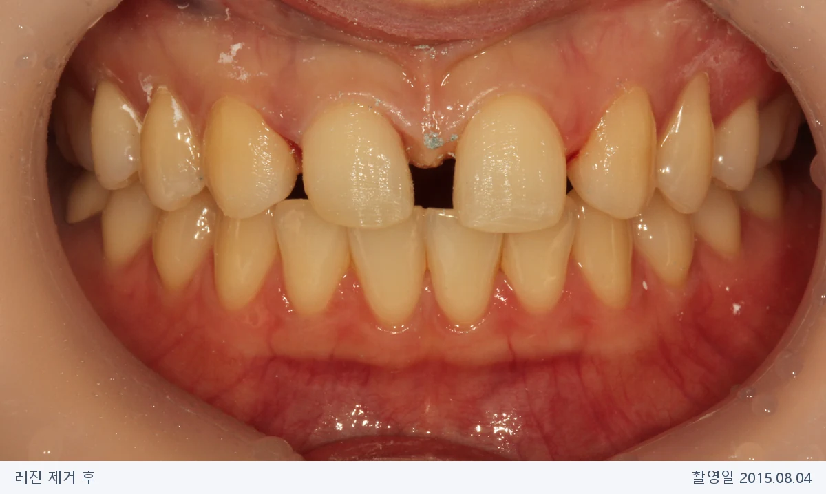
The original record describes resin filling the front-tooth spaces, with concerns about shape and recurrent gaps. Removing the resin revealed a large space between the central incisors.
Widening just these two teeth to fill the entire space could distort their proportions. We chose to move teeth orthodontically to distribute the spaces, then design restorations for those positions. Several months of tooth movement preceded veneer treatment.
The aim was not to place a veneer into an empty tooth site. It was to reposition the remaining teeth and reshape canines occupying lateral-incisor positions to suit the neighboring front teeth.
5. Treatment Stages by Photo Date
STEP 01
Assessing existing resin and proportions
The front spaces had been restored with resin despite both lateral incisors being absent. We assessed widths and overall alignment as well as gaps.
STEP 02
Viewing the spaces after resin removal
The resin was removed on the same day. The central space was assessed and orthodontic repositioning planned.
STEP 03
Distributing spaces through orthodontics
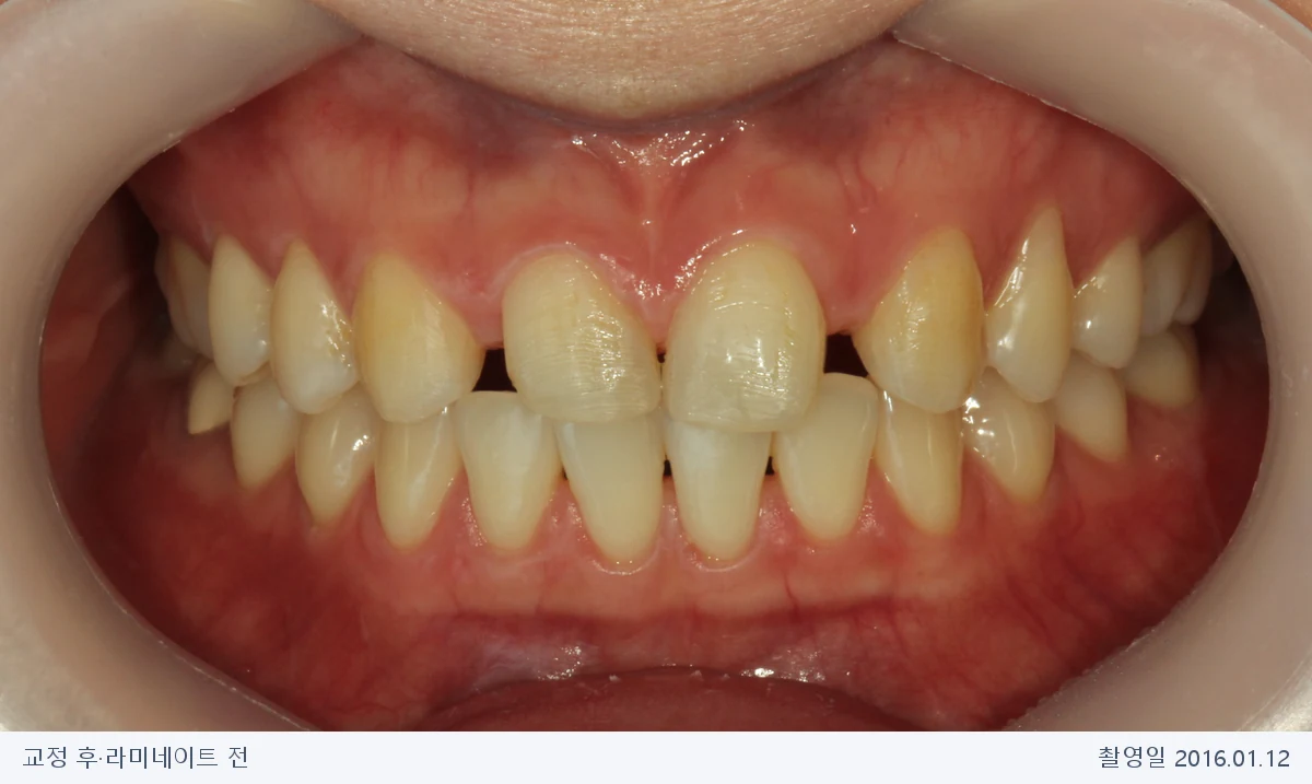
After orthodontics, before veneers, the spaces were distributed to allow suitable front-tooth contours rather than concentrated centrally.
STEP 04
Refining contours with veneers
The original clinical account records veneer fitting on the day the braces were removed. Canine contours were adjusted to resemble lateral incisors, alongside front-tooth proportions.
The two photographs dated 2016.01.12 show before and after fitting on the same day. Orthodontic duration, restoration extent and same-day suitability depend on tooth condition and planning.
6. Reviewing Shape and Retention after Treatment
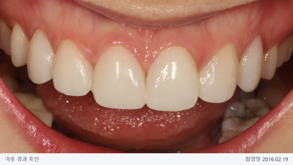
Veneer care includes later bite and retention checks as well as the fitting-day contours. Frontal and palatal photographs were taken on 19 February 2016. A retainer can be seen behind the front teeth.
These follow-up photographs were taken in February 2016.

7. Front-tooth Proportions within the Smile
Teeth can look different in close-up and within the smile. Before and follow-up smile photographs show their relationship to the lips. Facial photographs are limited to the mouth and chin area to reduce identifiability.
Lighting, angle and expression differ between dates. Tooth color and shape were not retouched; only crop boundaries, size and compression were adjusted. Results vary between patients.
8. Choosing Orthodontic Space Closure, Implants or Bridges
Treatment for missing lateral incisors can broadly involve orthodontic space closure and reshaping the remaining teeth or creating space to replace the missing teeth with restorations. Growth, alignment, the bite and canine size, shade and shape inform the plan.
Even with space closure, composite versus veneers is a separate restorative decision. Implants and adhesive bridges also require assessment of growth, gums and bone. This case’s sequence is not a universal plan for every missing lateral incisor.
The literature emphasizes assessment of tooth form and the bite, and collaboration between orthodontics and restorative care when canines substitute for lateral incisors. Read about assessment and canine substitution for missing lateral incisors
9. Aftercare and Points to Consider
- Orthodontic risks: Pain and gum inflammation may follow appliance fitting or adjustment; cleaning affects the risk of demineralization and decay. Recession or root resorption can occur, so monitoring is important.
- Retainer care: Follow the retention plan. If the appliance detaches or a gap appears, seek review rather than adjusting it yourself.
- Gums and restoration margins: Brush with fluoride toothpaste and clean between teeth. Veneers do not prevent decay around the restored teeth.
- Fracture and debonding: Avoid biting hard objects and discuss clenching. Wear, fracture or changes in bonding may require repairs or retreatment.
- Tooth preparation: Veneers may require removal of tooth tissue, which is irreversible. Not every case can be treated without preparation.
- Regular checks: Have the bite, gums, restoration margins and retainers reviewed. Discuss any further adjustments to shape or function when needed.
The American Dental Association’s veneer care guide also explains irreversible preparation, fracture risk and ongoing oral care.
For orthodontic risks and retention, see the British Orthodontic Society’s patient-information guidance. Discuss individual risks and alternatives with the treating clinician after examination.
10. Questions about Missing Front Teeth
Q. Are the teeth congenitally absent? How is an unerupted permanent tooth assessed?
Clinical and radiographic assessment is needed to determine whether a tooth never formed. If a permanent tooth has not appeared or the tooth count seems low, the cause should be assessed first. NHS information on congenitally missing teeth offers further explanation.
Q. Can missing front teeth be treated without implants?
For some missing lateral incisors, orthodontic space closure with canine reshaping is an option. Others are better served by space opening and implants or bridges. This case used orthodontics followed by veneers; suitability depends on growth, bite and tooth form.
Q. Can a veneer fill a missing-tooth space?
A veneer bonds to a remaining tooth surface. This case moved remaining teeth into appropriate positions and reshaped them, rather than placing veneers alone in empty spaces.
Q. Why align the teeth instead of filling the central gap directly?
Large or unevenly distributed spaces may produce unsuitable proportions if closed only by widening teeth. We assess whether position and bite need adjustment before deciding restoration extent. Missing teeth call for assessment of all remaining positions and spaces.
Q. Can a canine be reshaped to look like a lateral incisor?
This case used veneers to adjust the repositioned canines to suit adjacent front teeth. Canine substitution requires assessment of size, color, shape and bite; composite or veneer treatment is a separate decision.
Q. Is filling gaps with resin a poor choice?
No. Composite can be appropriate depending on gap size, tooth positions, bonding conditions and maintenance. The old restorations in this patient do not represent all composite treatment outcomes.
Q. Can veneers be fitted on the day braces are removed?
The original clinical account records this sequence for this patient. Actual timing depends on gums, restoration readiness and bite checks.
Q. Do veneers replace retainers?
No. Retainer type and duration follow the treating clinician’s plan. Detachment or deformation needs review.
Q. When were the photographs in this case taken?
The photographs document August 2015 to February 2016 and are presented with their treatment stages.
11. What Treatment Sequence Suits Your Teeth?
We assess missing-tooth positions, spaces, proportions and the bite together when discussing options.
12. References
- Alkadhimi AF, et al. Camouflaging the permanent maxillary canine to substitute an absent lateral incisor – part 1: assessment and management. British Dental Journal. 2022;232:20–26. PubMed article information
- American Dental Association. Veneers. MouthHealthy. Patient information (accessed 2026.10.17)
- British Orthodontic Society. Risks of Orthodontic Treatment: Guidance on Informing Patients. Reviewed 2023. Orthodontic risk guidance (accessed 2026.10.17)
These references provide general information about treatment selection and care.
Treatment and original clinical account: Dr. Hanna Lee, Harue Dental
Clinical photo dates: 2015.08.04, 2016.01.12, 2016.02.19
Text and photo editing: 2026.10.17
Photos and clinical information are published within the patient’s consent for website use. This is one patient’s case. Methods, duration and results depend on the individual teeth and gums; a precise plan requires examination.